The spider veins, also known in the medical world as telangiectasias, are an aesthetic concern frequent. These tiny dilated veins, although generally harmless, can significantly affect your self-esteem if you suffer from them.
In it García-Legaz Dermatological Institute, we want to delve into what spider veins are, factors that contribute to its formation and the available treatments to achieve a more even complexion.
What are spider veins?
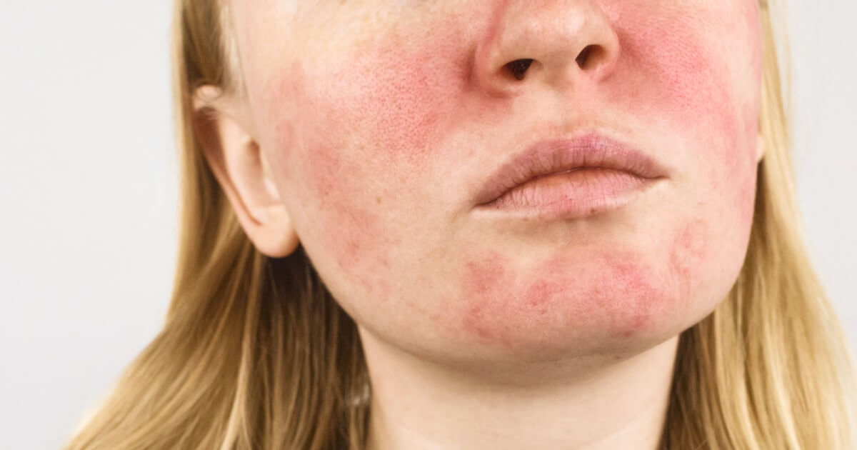
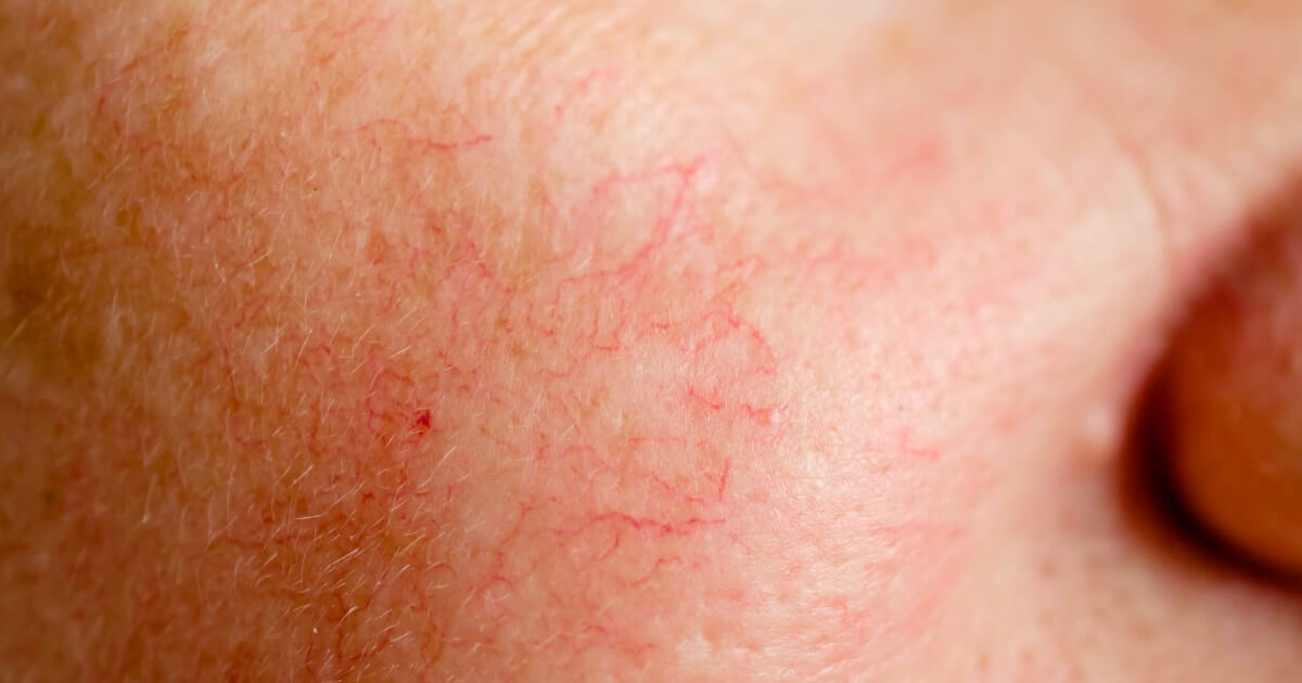
Spider veins are dilations of small blood vessels that are made visible through the skin. They manifest as fine reddish, bluish, or purple lines, forming patterns similar to cobwebs, which explains its colloquial name.
"The telangiectasias, usually asymptomatic, can range in size from just a few millimeters to several centimeters in diameter.”
Although they can appear anywhere on the body, they are more common in the legs and the face, especially on the cheekbones and nose.
Unlike varicose veins, which are more prominent and bulging, spider veins are characterized by being flat and, generally, they do not cause physical discomfort. However, his aesthetic impact can be considerable, especially when they appear in visible areas.
How spider veins form
The development of spider veins is a process complex that can be influenced by several factorsAt the García-Legaz Dermatological Institute, we mainly identify the following: causes:
- Genetic predisposition: if you have family history of telangiectasias, you are more likely to develop them.
- Hormonal changes: Conditions such as pregnancy can trigger the appearance of spider veins. During pregnancy, the increased blood volume and the hormonal changes They can dilate blood vessels, increasing their visibility.
- Sun exposure: Ultraviolet radiation can weaken the walls of the vessels superficial blood vessels, making them more susceptible to dilation. In addition, prolonged exposure without proper protection can exacerbate existing telangiectasias.
- Blood pressure: conditions that increase the pressure in the veins, such as standing or sitting for long periods, can promote the appearance of spider veins, especially in the legs.
- Aging: over time, the fur becomes more fine and the blood vessels become more visible. Also, the dnatural deterioration of venous valves can contribute to their formation.
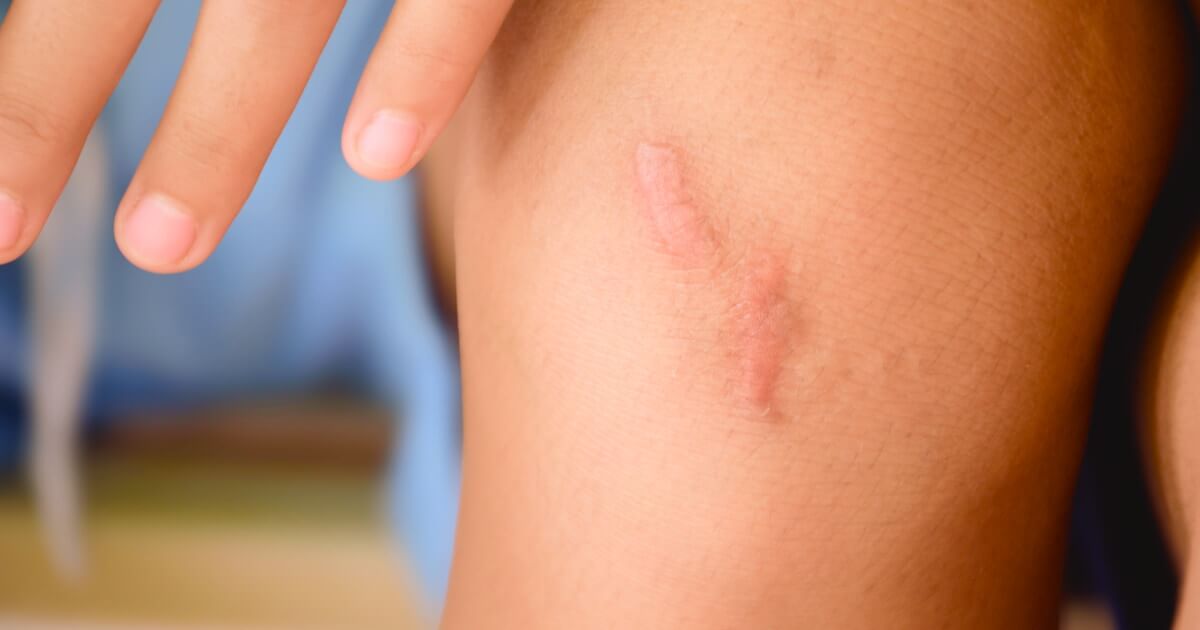
- Skin lesions: trauma, even minor ones, can cause localized dilation of the blood vessels.
How do you remove spider veins?
Fortunately, dermatology has various effective treatments for spider veins. However, choosing the most suitable one for you will depend on factors such as location and extension of spiders, as well as of you skin type, medical history and preferences personal.
Below, we discuss the most common and effective options:
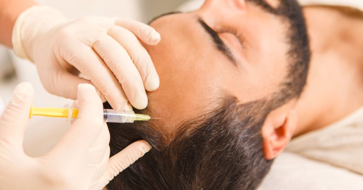
Sclerotherapy
Sclerotherapy is a standard treatmentr for the removal of spider veins.
This technique consists of inject a sclerosing solution directly into the affected veins. This substance causes a controlled irritation inside the vein, causing its collapse and later reabsorption naturally by the body, considerably reducing its appearance.
Vessels smaller than 1 mm in diameter are difficult to inject and remove in a single session, so they are generally needed. various treatments to eliminate them completely.
After the procedure, it is common for a mild redness, bruising, or swelling in the treated area, although they usually disappear in a short time.
Laser treatment of spider veins
Laser treatment is an option highly effective to remove spider veins, especially in delicate areas such as the face. In addition, at the García-Legaz Dermatological Institute we use the advanced Nordlys platform, which ensures maximum precision and minimizes side effects.
“The dermatological laser has the ability to treat small and medium caliber blood vessels, with visible results from the first session.”
This technique non-invasive uses a beam of light that is selectively absorbed by the hemoglobin of the blood vessels. The light generates heat, causing the coagulation and the closing of the vessels without damaging the surrounding skin.
As these vessels collapse, the body naturally absorbs them, leaving the skin with a more uniform appearance and significantly improving the appearance of spider veins.
Post-treatment care
To maximize the results of any spider vein treatment and minimize potential side effects, it is essential to follow certain care guidelines:
- Photoprotection: use daily sunscreen Broad spectrum with SPF 50 to prevent the appearance of new spider veins and protect treated skin, even on cloudy days.
- Hydration: Keep your skin well hydrated to promote a optimal recovery and prevent the appearance of new visible blood vessels.
- Avoid sun exposure: especially in the first few weeks after treatment. If being outdoors is unavoidable, use protective clothing and look for areas of shade.
- Following medical instructions: If your dermatologist recommends wearing compression stockings or other measures, be sure to follow them. maximize results.
- Healthy lifestyle: keep a adequate weight, beam exercise regularly to improve circulation and avoid stay in the same position for extended periods.
- Patience: The final result may take several weeks to arrive, so it is important to be constant and follow the recommendations from your dermatologist.
In conclusion, there are effective treatments for spider veins. Our specialists in dermatology They will evaluate your particular case and they will design a treatment plan tailored to your specific needs, using the more advanced and safer techniques to help you regain even-looking skin.
Literature
- Hadad, IP (1999). Laser treatment of cutaneous vascular lesions. Journal of the Colombian Association of Dermatology and Dermatologic Surgery, 7(2), 65-69. https://www.revista.asocolderma.org.co
- Goldman, M.P., & Bennett, R.G. (1987). Treatment of telangiectasia: a review. Journal of the American Academy of Dermatology, 17(2), 167-182. https://www.sciencedirect.com
- Arellano, A., & Ríos, L. (2005). Treatment of leg telangiectasias with laser and high-intensity pulsed light. Cosmetic, Medical and Surgical Dermatology, 3(2), 83-86. https://www.medigraphic.com